
6X Tooth Decay Pathological Comparison Model

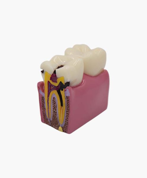
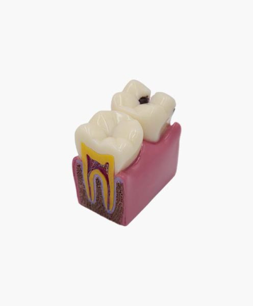
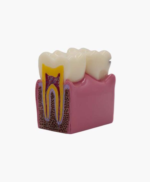
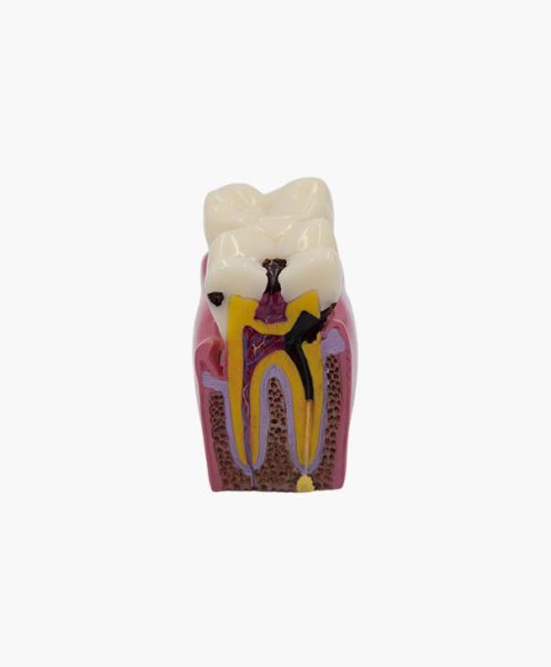
6X Tooth Decay Pathological Comparison Model
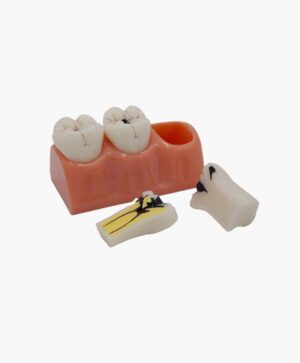
- Normal pulp, nerves, vessels can be seen as well as pulp lesions from deep caries
- Used for teaching caries and endodontic lesions
Additional information
| Material | Resin |
|---|---|
| net weight | 302g |
| gross weight | 340g |
| Packaging size | 12*7.6*10cm |
Related Products
Periodontal Classification Model (Soft Gingiva)
Periodontal Classification Model (Soft Gingiva)
Available:
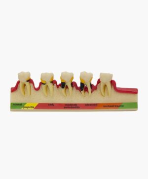
Periodontal Classification Model (Soft Gingiva)
Periodontal Classification Model (Soft Gingiva)- Demonstrates pathological status at different stages of periodontal disease and alveolar bone resorption due to occlusal trauma
- With soft gingiva
- Used for teaching periodontology
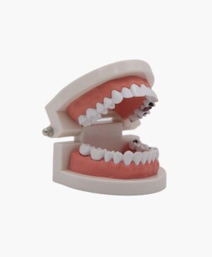
Small Pathological Tooth Model
Small Pathological Tooth Model- Small tooth decay model
- For children’s pathological tooth decay teaching
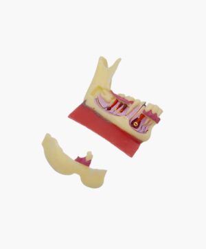
Mandibular Tissue Detachable Model
Mandibular Tissue Detachable Model- Normal size mandible with pathological changes
- Part of the jaw can be opened to see inside the nervous system , tooth root, purulent pulp infection
- Used for teaching of pathological changes of jaw bones
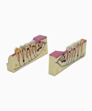
2 Times Right Lower Tooth Detachable Model
2 Times Right Lower Tooth Detachable Model- Visible nervous system, tooth root, crown with inlay, amalgam filling, abscess, low impacted wisdom tooth, local inflammatory reaction
- Can be used for teaching endodontics and restoration
4X Tooth Decay Model
4X Tooth Decay Model- 3 movable longitudinally sectioned teeth represent the 3 stages of caries
- Used for teaching caries with different levels

Standard Pathological Missing Tooth Model
Standard Pathological Missing Tooth Model- Can be used for orthodontic simulation training
Reviews
There are no reviews yet.